French Bulldog Eye Ulcer: Signs, Treatment & 2026 Costs
TL;DR
- A squinting Frenchie is a French Bulldog eye ulcer until a vet proves otherwise. An eye held shut, watery, or cloudy needs a same-day call, not a wait-and-see weekend.
- French Bulldogs had 7.25 times the odds of corneal ulcer disease compared with crossbreds in VetCompass data, with a one-year prevalence of 1.87 percent. One of the highest-risk breeds, though Pugs ranked higher.
- An ulcer means the cornea’s surface layer is gone and the damage has reached the middle layer, the stroma. Deep ulcers can worsen overnight and are emergencies.
- Put the cone on and keep it on. Never use steroid drops on an ulcerated eye, or leftover or human drops without your vet’s approval.
- A simple ulcer caught early often totals a few hundred dollars. The stain averaged $36 in US data, treatment averaged $677 across all severities, and PetMD quotes surgery at $600 to $2,000.
- Healing is proven by a repeat stain at a recheck, not by the eye looking better. See our French Bulldog health problems hub for the breed’s other common problems.
He Went to Sleep Fine and Woke Up Squinting
Your French Bulldog woke up with one eye clamped shut, tears running down the fold under it, pawing at his face on the rug. In this breed, that picture points first to a corneal ulcer: a raw spot where the clear front surface of the eye has lost its outer layer. The cornea is rich in nerves, and dogs with ulcers rub the eye with a paw or along the carpet.
An ulcer starts as damage to the epithelium, the cornea’s thin outer layer. A scrape that stays in that layer is an abrasion. Once the erosion breaks through into the stroma beneath, vets call it an ulcer. Deeper still and only Descemet’s membrane remains, a state called a descemetocele. If that membrane ruptures, fluid leaks out and the eye can collapse. VCA Animal Hospitals lays out that three-layer anatomy, and why depth, more than size, decides urgency. Most ulcers start with injury: a nail scratch, a thorn, shampoo at bath time, or a hair rubbing daily. In a Frenchie, the breed’s own anatomy often does the rubbing.
Quick tip: Photograph both eyes today, same light, and again tomorrow. Dated photos show whether the cloudy spot is growing.
Why a French Bulldog Eye Ulcer Happens So Easily
In O’Neill et al. 2017, a VetCompass study of 104,233 dogs under primary care in England, French Bulldogs had 7.25 times the odds of corneal ulcerative disease compared with crossbreds. Eye disorders hit 10.5 percent of French Bulldogs in the breed-specific VetCompass study, and ulcerative keratitis alone accounted for 2.1 percent.
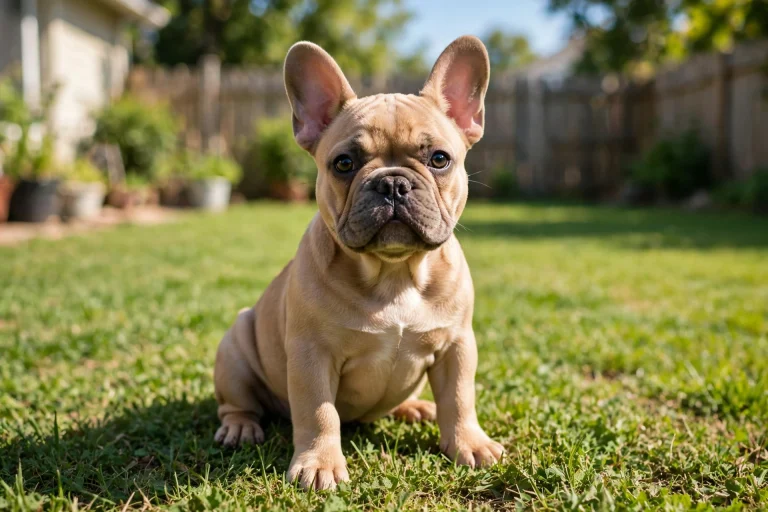
Frenchies have shallow eye sockets, so the eyes sit proud of the skull instead of tucked deep in bone, a trait called exophthalmia. A protruding eye meets the world first: the cat’s paw, the rose bush, the coffee table edge. Packer, Hendricks and Burn tied flat-faced conformation directly to ulceration, and found dogs with a nasal fold carried nearly five times the risk, because fold hairs rub the cornea with every blink, a fault called nasal fold trichiasis.
Many Frenchies cannot close their lids fully, a gap called lagophthalmos, so the cornea dries between blinks and during sleep. Three quieter faults complete the picture:
- Medial entropion: the lower lid’s inner corner rolls inward, often hidden under the nose fold, so lashes drag across the cornea.
- Distichiasis and ectopic cilia: extra or misplaced lashes touch the eye. Ectopic cilia poke through the lid lining and can cause intense pain and ulcers.
- Reduced corneal sensitivity: central sensitivity is reported 50 to 93 percent lower in brachycephalic dogs than in longer-nosed dogs, so an early ulcer may not trigger the blinking that warns an owner.
The tear film adds a final twist. In healthy French Bulldogs, a 2025 study found tear volume was actually fine, but the film broke up in half the normal time: 7.39 seconds against 14.74 in non-brachycephalic controls.
Signs of a French Bulldog Eye Ulcer: What You Will See
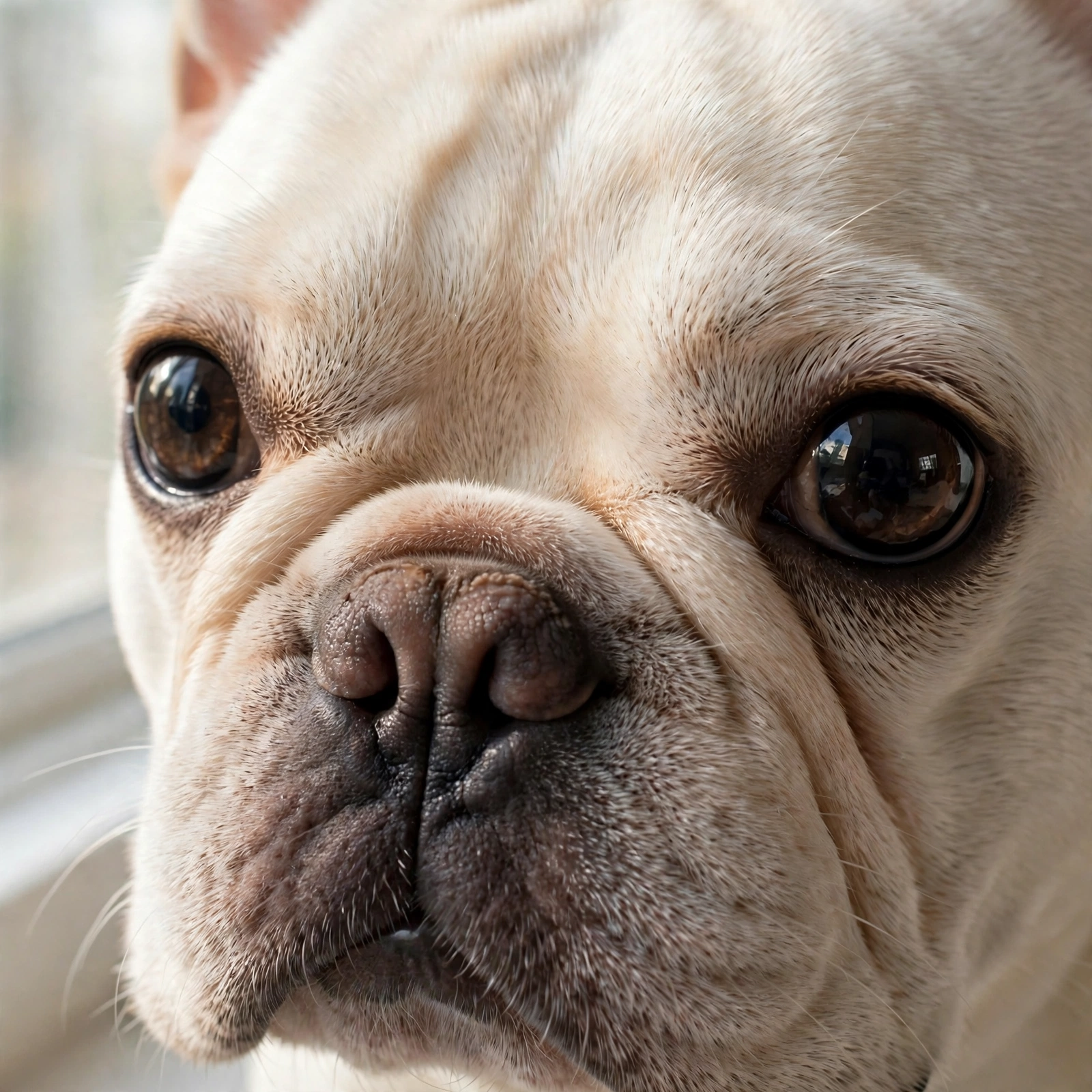
Pain shows first. A Frenchie with an ulcer squints, blinks hard, or holds the eye shut. The white of the eye turns red and clear tears spill over. Many dogs paw at the face, and the third eyelid often rises partway across the eye.
Fluid soaks into the stroma where the surface is missing, so the area looks cloudy or blue-grey. A dense white or yellow patch suggests white blood cells and bacterial involvement. Yellow or green discharge, or a dog who stops eating, needs a same-day call.
| What you see | What it often means |
|---|---|
| Squinting, eye held shut, pawing at the face | Pain, ulcer high on the list |
| Cloudy or blue-grey spot on the eye | Fluid in the stroma at an ulcer site |
| Yellow-green discharge | Infection, see a vet the same day |
| A visible dent or crater on the eye surface | Deep ulcer, emergency |
| Smooth pink lump at the inner corner | More likely cherry eye than an ulcer |
One warning deserves its own line. Eye Vet UK specialists note that a deep ulcer can look clearer and seem less painful as it sinks past the surface nerve endings. Quieter can mean closer to rupture, not healing.

The Types, From Surface Scratch to Emergency
Vets sort ulcers by depth and behaviour, and the label decides treatment:
| Type | What is damaged | How it behaves |
|---|---|---|
| Superficial ulcer | Epithelium only, or the top of the stroma | Heals in about a week with drops and a cone |
| Indolent ulcer, called SCCED | Epithelium that will not stick to the stroma beneath | Lingers for weeks until the loose edge is removed |
| Stromal or deep ulcer | Well into the stroma, usually with bacterial infection | Bacteria release enzymes that digest the cornea deeper |
| Melting ulcer | Stroma dissolving under enzyme attack | Soft, grey-yellow, gelatinous look. Emergency care |
| Descemetocele | Stroma gone, only the last membrane left | A crater with a clear base. Surgical emergency |
| Perforation | The membrane has ruptured | Fluid leaks, iris may plug the hole, vision at risk |
Superficial ulcers heal fast because new surface cells start sliding across the defect within an hour. VCA puts abrasion healing at 3 to 5 days and PetMD puts simple ulcers at 7 to 10 days, so plan on roughly a week and a recheck stain to prove closure. The indolent ulcer fails mechanically: a thin membrane forms over the stroma and fresh epithelium cannot grip it, so it will not heal until a vet debrides the loose edge.
Ulcers deeper than about half the corneal thickness generally need surgical support and immediate referral to a veterinary ophthalmologist, not drops alone. A melting ulcer, driven most often by Pseudomonas, Staphylococcus, or Streptococcus, can worsen overnight, so vets recheck within a day and often hospitalise the dog.
How Vets Diagnose an Eye Ulcer
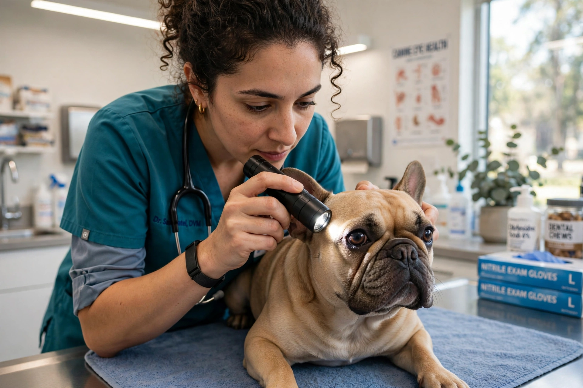
The vet places fluorescein, an orange dye, on the eye. It washes off a healthy surface but sticks to exposed stroma, so an ulcer lights up green under a blue light: the dim room, the blue lamp, a bright green patch where the damage sits. PetMD’s ulcer guide describes the same test as the standard first step, with one quirk. A descemetocele takes stain only around its rim, because the membrane at its base will not absorb the dye. A clear centre ringed in green is bad news, not a clean result.
Tear testing comes early, ideally before any drops. For the Schirmer tear test, a paper strip sits inside the lower lid for one minute. Normal is above 15 mm per minute, and under 10 points to clinical dry eye. Your vet may add tonometry, a bacterial culture swab, or a cytology sample, especially for a deep, melting, or non-healing ulcer.
Checklist: what to tell the vet
- When the squinting started, over hours or days
- Any rough play, cat contact, brush, or bath in 48 hours
- Every drop or ointment already used, bottles in hand
Referral is routine for the serious end of the ladder. Melting, deep stromal, and complicated ulcers usually go to a veterinary ophthalmologist. Complicated cases are rechecked in 1 to 2 days, simple ulcers in 5 to 7, and if healing stalls, VCA advises re-examination after 2 to 3 days.
Treatment, Step by Step
Treatment follows a ladder. Most simple ulcers never pass the first rungs.
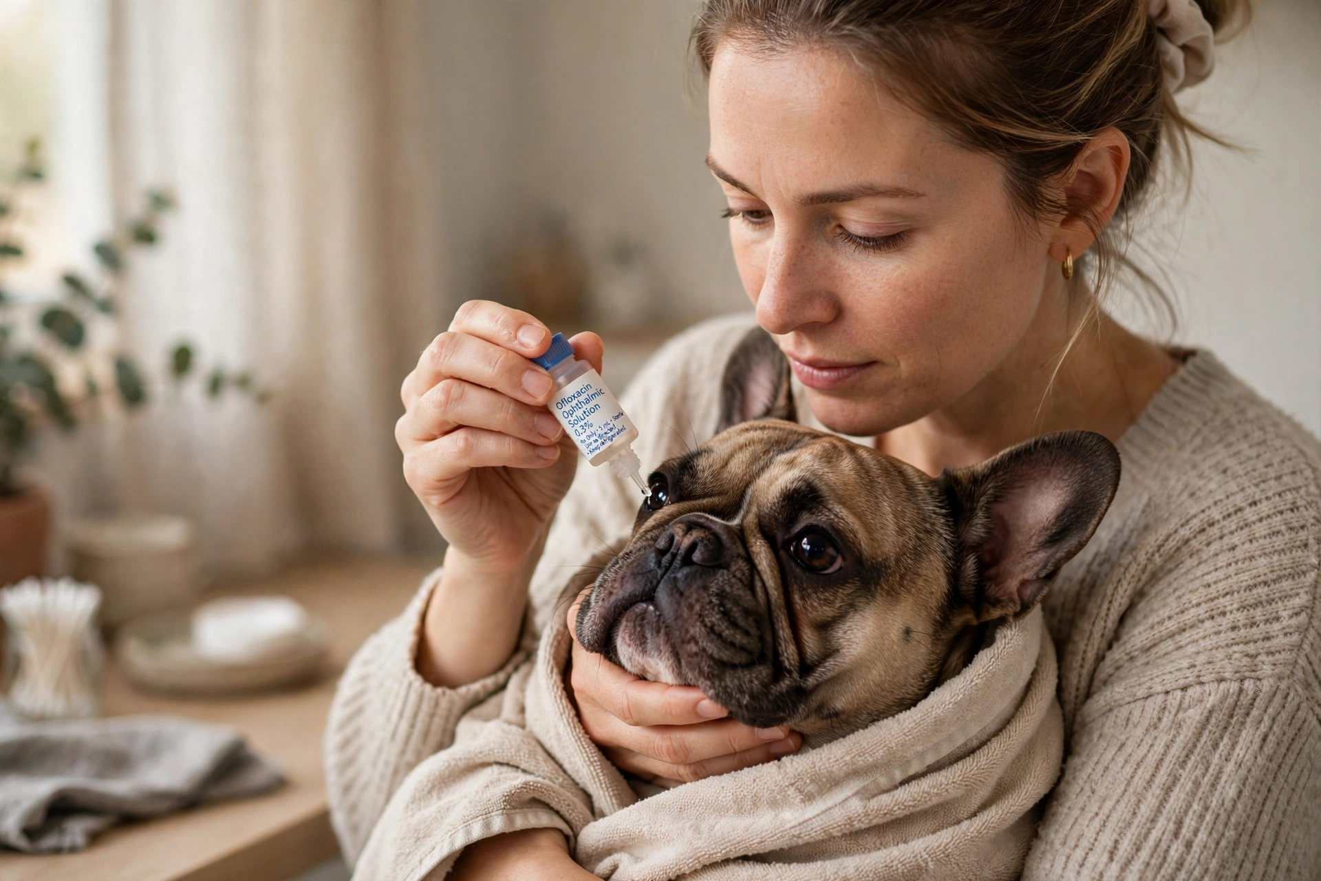
- Topical antibiotics. Drops guard the open cornea against infection while it heals, but they wash away quickly, so VCA advises applying them every 4 to 6 hours in typical cases. Ointments last longer between doses. Set phone alarms, including one overnight alarm.
- Atropine. This drop relaxes the muscle inside the eye that spasms when the cornea is injured, and dilates the pupil. Dilation and light sensitivity can last several days, so expect a big black pupil and squinting at the patio door. A little drains into the throat and tastes intensely bitter. Drooling and pawing at the mouth after this drop is a taste reaction, not an allergy.
- Oral pain relief. Many vets add an oral anti-inflammatory such as carprofen. Topical anaesthetic drops are a one-time diagnostic tool at the clinic only. Repeated at home, they damage the cornea and delay healing.
- Serum drops. For deep or melting ulcers, the vet may spin down a sample of your dog’s own blood and use the serum as drops. Serum carries nutrients and anti-collagenase activity that slows the enzymes dissolving the stroma. Keep the bottle refrigerated.
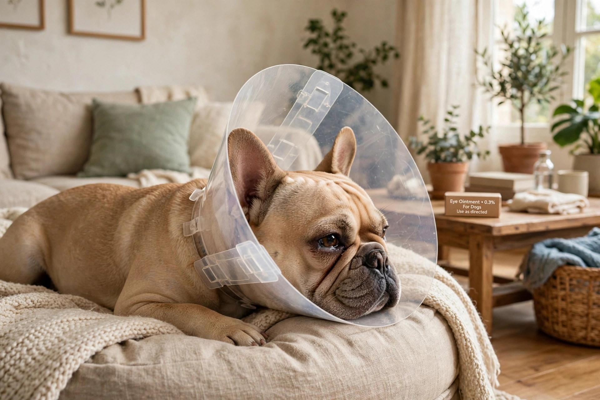
- The cone. Every step above fails if a paw reaches the eye. VCA is blunt: the dog must wear an Elizabethan collar to stop rubbing. One good rub can strip off new surface cells that took days to grow.
Never stop treatment early because the eye looks calm again. Continue until the vet repeats the stain and confirms closure. Rechecks run every 1 to 7 days by severity, and red streaks creeping toward the ulcer are new blood vessels, a normal healing sign.
When Drops Are Not Enough: Procedures and Surgery
Two situations push a case beyond drops. The first is the indolent ulcer that will not stick. The fix is removing the loose surface so fresh cells can grip. Referral centres report these success rates:
- Cotton-swab debridement alone: about 50 percent heal
- Grid keratotomy: about 60 to 70 percent
- Diamond burr debridement: roughly 7 to 9 in 10 in referral reports
- Superficial keratectomy under general anaesthesia: about 95 to 100 percent
Treat those as centre estimates, not guarantees. Some dogs need a second procedure, and the cone matters even more after debridement, while neighbouring cells are still poorly attached.
The second situation is depth. For a deep ulcer, a descemetocele, or a perforation, the surgeon removes dead tissue and gives the cornea structure and blood supply. The usual answer is a conjunctival graft, a flap of the pink tissue lining the lid, stitched over the defect. Corneal grafts and third-eyelid flaps are other specialist options. Merck notes that many deep ulcers require a conjunctival graft, and PetMD notes these grafts usually stay in place for 2 to 3 months.
Whatever the procedure, the cause has to be fixed in the same plan. An ulcer over an in-rolled lid or an ectopic cilium returns until the lid is corrected or the hair removed. Ask the surgeon what caused this ulcer and what stops the next one.
Do this, not that:
- Do ask for the referral early if the ulcer is deep or melting. Do not spend a week cycling drops while the cornea thins.
- Do expect drops every 2 to 4 hours in a hospitalised melting case.
What It Costs in the US in 2026
No national fee survey covers corneal ulcer care, so read these as sourced ranges, not quotes. Emergency and big-city prices run higher.
| Step | Typical 2026 US figure |
|---|---|
| Vet exam | $50 to $150 |
| Fluorescein stain test | Average $36, range $28 to $71 |
| Eye medication, per course | About $20 to $100 or more |
| Oral pain medication | About $15 to $60 or more |
| Recheck exam | $30 to $100 or more |
| Ophthalmologist consultation | $150 to $300 or more |
| Eye ulcer surgery, per PetMD | $600 to $2,000 |
Both figures come from the Synchrony and CareCredit 2024 procedural cost study, the strongest US dataset found for this condition. Its $677 figure averages the cost of treating a corneal ulcer across all severities, medical and surgical cases mixed, range $520 to $1,306. It is not a surgery price.
A straightforward superficial ulcer seen promptly commonly totals a few hundred dollars: exam, stain, drops, and one recheck. An indolent ulcer needing a grid or diamond burr procedure, or a deep ulcer needing a conjunctival graft, moves into the $600 to $2,000 surgery range PetMD quotes, and complex referral or emergency graft cases are quoted at $1,500 to $3,000 or more in 2026 cost guides. No authoritative US price list separates grid keratotomy from diamond burr work, so both sit inside the surgery ranges above.
Home Care Rules That Protect the Eye
Home care is where ulcers are won or lost. The rules fit on the fridge door.
Do this, not that:
- Do keep a hard plastic cone on 24 hours a day until the vet clears it, fitted so the rim extends past the nose. Do not swap in a soft inflatable collar. A Frenchie can fold those and reach the eye.
- Do give every drop on schedule and finish the course. Do not stop because the eye looks better on day four.
- Do call the same day if squinting returns, discharge turns yellow-green, or the cloudy area grows.
Now the rule that protects the eye most. Never use steroid drops on an ulcerated eye. VCA warns that corticosteroids will slow or stop healing and may cause serious complications, and steroid use is a listed risk factor for melting ulcers. Check any leftover bottle before it goes near the eye. Look for prednisolone, dexamethasone, or hydrocortisone on the label, including short forms like pred, dex, or HC, and combination names such as Neo-Poly-Dex. Use nothing else in that eye without your vet’s say-so. Steroids enter the picture only after a repeat stain confirms closure, and if the eye becomes painful again on one, VCA advises stopping and rechecking at once.
Expect the cone to clatter against doorframes and water bowls for a week or two. Raise the bowl, clear a cone-width path to the door, supervise stairs, and fill the hours with frozen food toys.
Other French Bulldog Eye Problems to Know
Ulcers are the emergency, but they share the breed with slower problems, several of which cause ulcers when ignored.
| Problem | What happens | The fix in brief |
|---|---|---|
| Dry eye, called KCS | Too little watery tear production, or a tear film that breaks up too fast. Dull cornea, thick discharge, repeat ulcers | Tear stimulants such as cyclosporine or tacrolimus, plus lubricants |
| Pigmentary keratitis | Brown-black pigment spreads across the cornea after chronic irritation | Treat the underlying irritation. Dense pigment usually cannot be reversed |
| Entropion | The lid margin rolls inward, in Frenchies at the inner corner and on both sides | Surgical correction of the lid |
| Distichiasis and ectopic cilia | Extra or misplaced lashes touch the cornea and keep an ulcer open | The hair must be removed. Plucking is temporary |
| Conjunctivitis | Inflammation of the eye lining, 3.2 percent of Frenchies in the VetCompass breed study | The common look-alike. Treated by cause, after a stain rules out an ulcer |
Corneal scarring, called fibrosis, is the footprint a healed ulcer leaves, seen in a quarter of one brachycephalic referral sample. With sockets this shallow, a hard blow can push the eye out of the socket. That emergency, proptosis, needs a vet within the hour and a moist, protected eye on the drive.
For the lump rather than the raw spot, see our guide to cherry eye in French Bulldogs. That gland produces a large share of the watery tear film, so its health ties straight back to dry eye and ulcer risk.
Can You Prevent Ulcers?
You can lower the risk. You cannot design it away, because the main risk factor is the shape of your dog’s head. These habits target the injuries that turn anatomy into an emergency:
- End games that put claws near the face, and supervise play with cats and boisterous dogs.
- Keep your Frenchie out of tall weeds, brush, and thorny hedges. Choose the mown path.
- Shield the eyes at bath time and rinse shampoo splashes with plain water at once. Chemical irritation is a named ulcer cause.
- Keep nose-fold hairs trimmed away from the eye, and raise hairs that touch the cornea with your vet.
- If your dog sleeps with lids parted, ask your vet about a bedtime lubricating ointment.
- Treat the drivers: lid surgery for entropion, lash removal for distichiasis, and daily dry-eye medication.
No diet, supplement, home eye wash, or natural drop was verified as preventing ulcers, so be wary of any product that promises it. Regular eye checks at routine visits are the prevention specialists recommend for brachycephalic dogs. Our complete French Bulldog owner’s guide folds those checks into the breed’s wider care routine.
Frequently Asked Questions
Will an eye ulcer heal on its own? A surface abrasion sometimes closes without treatment, but an owner cannot tell an abrasion from an ulcer at the kitchen table, and a true ulcer left alone can deepen and scar. Vets treat a squinting Frenchie as an ulcer until a stain proves otherwise.
How long does an eye ulcer take to heal? A simple superficial ulcer usually closes in about a week, with published ranges of 3 to 5 days to 7 to 10 days. Indolent ulcers take weeks and need a procedure. Deep ulcers run longer, with rechecks every 1 to 2 days and grafts left in place for 2 to 3 months.
Is an eye ulcer an emergency tonight? Squinting with a cloudy spot, a visible dent, yellow-green discharge, or an eye held shut after a scratch means tonight, not Monday. A deep or melting ulcer can worsen overnight. Mild redness with a comfortable, open eye can usually take the first morning appointment.
Why is my dog drooling after the eye drops? That is usually the atropine. A little drains into the back of the throat, and it tastes intensely bitter. Drooling for a few minutes after the dose is a taste reaction. Call sooner if it lasts for hours.
Can an ulcer come back, or affect the other eye? Yes to both. In the VetCompass data, 5.4 percent of cases recurred in the same eye, and the anatomy behind the first ulcer is present in both eyes. An ulcer from an in-rolled lid or a stray lash keeps returning until that cause is fixed.
Can I use my own eye drops, or drops left over from another problem? No. Human and leftover pet drops can contain steroids, and steroid drops on an ulcer slow healing and raise the risk of melting. Even plain lubricating tears should get a quick phone approval first.
Final Word
A French Bulldog eye ulcer is painful, common in this breed, and very treatable when the first squint earns a same-day call, not a watchful weekend. Keep the cone on, guard the drop schedule, and let a green stain at the recheck tell you when the eye has truly closed. Save this guide for the first time those big eyes look cloudy, and send it to the Frenchie owner who texts you a one-eyed photo. Those eyes meet the world first. Your job is to get help fast.