Cherry Eye in French Bulldogs: Treatment & Surgery Costs
TL;DR
- Cherry eye in French Bulldogs is a prolapsed tear gland of the third eyelid: a smooth pink-red lump at the inner corner of the eye. It is common in the breed, and it is not a tumor.
- The gland produces roughly a third to a half of the watery tear film, which is why vets tuck it back into place instead of removing it.
- Surgical repositioning is the standard fix. Performed promptly in a young dog, veterinary ophthalmologists report it is almost 100 percent effective.
- In 2026, most US owners pay roughly $500 to $1,500 per eye at a general practice vet, or $1,200 to $2,500 at a specialist. Fixing both eyes at once typically runs $800 to $2,500.
- Do not massage it back yourself, and do not wait it out. About 43 percent of untreated cases develop dry eye, and chronic cases are harder to fix.
The Red Lump in the Corner of the Eye
You were rubbing your Frenchie’s belly when you spotted it. A round, cherry-red lump tucked into the inner corner of his eye, right where it meets the nose. It was not there yesterday. Your stomach dropped, because the first word that surfaced was tumor.
It is almost certainly not a tumor. What you are looking at is cherry eye, and in French Bulldogs it ranks among the most common eye problems a vet will see. This article explains what that lump really is, why this breed gets it so often, what the vet will actually do, what it costs in 2026, and which mistakes turn a fixable problem into a lasting one.
Quick tip: Snap a clear photo of the lump today. Early cherry eye can pop in and out, and a photo proves what you saw if it vanishes before the appointment.
What Cherry Eye in French Bulldogs Actually Is
Dogs have a third eyelid, a thin membrane tucked just inside the lower lid that sweeps across the eye with every blink. At its base sits a small tear gland wrapped around a T-shaped piece of cartilage. Cherry eye is that gland slipping out of position and bulging into view as a smooth pink-to-red mass at the inner corner. Vets call it prolapsed nictitating membrane gland, usually shortened to PNMG.
The nickname comes from the look. Once the gland pops out, it swells into a round red lump that genuinely resembles a cherry.
Here is why the gland matters. It makes roughly a third to a half of the watery part of the tear film. The American College of Veterinary Ophthalmologists puts the share at 33 to 66 percent, while VCA Animal Hospitals says up to 50 percent. That single fact drives every treatment decision in this article. A gland responsible for that much of the eye’s moisture is worth saving, not cutting out.

Quick tip: The lump often looks worse than it is. A smooth, round, pink mass at the inner corner is the textbook picture. A jagged, bleeding, or rapidly growing growth is a different story and needs urgent care.
Why French Bulldogs Get It So Often
The short answer is anatomy. The gland is normally anchored to the lower inner rim of the eye by a fibrous attachment, and in certain breeds that anchor is weak. The precise cause remains unknown, but veterinary ophthalmologists describe it as possibly multifactorial: not just a loose ligament, but a function of how the whole head is built in Bulldogs. Crowding of the lower eye socket in flat-faced breeds is one suggested contributor, and a 2024 genetic study linked a gene variant to higher risk. That variant is a risk factor, not destiny.
The numbers match what Frenchie owners observe. In the largest study of its kind, covering more than 900,000 UK dogs, brachycephalic breeds had 6.71 times the odds of cherry eye compared with longer-nosed breeds. The French Bulldog was the third most common breed among the 1,802 cases, and VCA lists the Frenchie among the most-affected breeds outright. You can read the full study in PLoS ONE.
It is also a young dog’s condition. The median age at first diagnosis was 0.63 years, about seven and a half months. Dogs under one year had 10.82 times the odds of dogs aged two to four, and most cases appear before age two. Early-onset cases can show up at two to three months old.
Quick tip: The research does not support a meaningful sex difference. The large UK study found sex itself was not associated with cherry eye odds, so ignore anyone who tells you males are the ones to watch.
Signs: What You Are Looking At
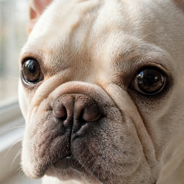
The classic sign is unmistakable once you know it. A smooth red or pink mass sits on the lower lid near the nose, at the inner corner of the eye. It may be large enough to cover part of the cornea, or small and only periodically visible. You might also notice extra tearing, a bit of mucus discharge, or mild swelling around it. If the eye feels irritated, the dog may rub at it or squint.
One reassuring fact: cherry eye does not usually hurt at first. Veterinary reviewers at WebMD put it plainly. It doesn’t usually hurt, but the dog might rub the area, irritate it, and make it bleed. Pain signals such as squinting, a cloudy eye, or thick discharge point to a complication, not to the cherry eye itself.
How to tell it apart from other red-eye problems:
| What you see | What it usually means |
|---|---|
| Smooth pink-red lump at the inner corner | Cherry eye |
| Redness spread across the whole eye | Conjunctivitis or an allergy flare |
| Squinting with a cloudy or bluish spot | Possible corneal ulcer, see a vet promptly |
| Thick yellow-green discharge | Infection, see a vet promptly |
Diagnosis happens by physical exam, which vets describe as fairly easy to recognize. Your vet may also run a Schirmer tear test, a small paper strip that measures tear production, and a fluorescein stain that reveals corneal ulcers under blue light. Both take minutes and cause no pain.
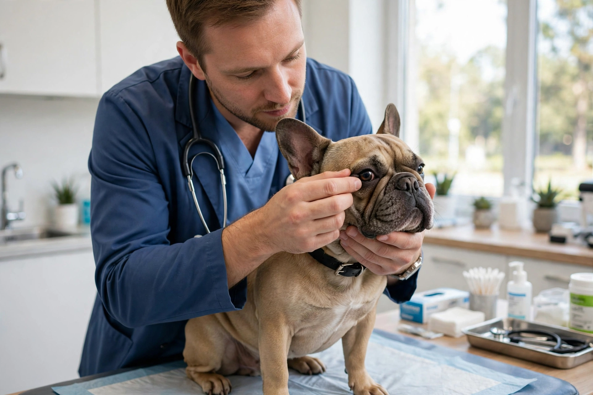
Quick tip: Try the smooth-versus-angry check at home. A cherry eye lump looks like a distinct rounded mass. An eye that is red all over with no distinct lump is a different problem and needs its own diagnosis.
Can You Massage It Back? The Honest Answer
This is the question every owner asks, usually after watching a video online. Here is the honest version.
A vet can sometimes press a recently prolapsed gland back into place, occasionally with numbing drops or light sedation, and pair it with anti-inflammatory medication. As a stopgap, that is legitimate medicine. But it is usually temporary. Most glands put back this way prolapse again, and manual tucking is widely described as rarely staying put.
What you should not do is try it at home. Pressing on an inflamed, exposed gland risks scratching the surface of the eye, and the gland is more delicate than it looks. The veterinary guidance is blunt. Don’t try to push the gland back yourself. When it is done, it is done by a vet, using numbing drops.
Do this, not that:
- Do call your vet promptly and ask for the soonest available appointment. Do not wait to see whether it resolves on its own.
- Do keep your dog from rubbing the eye. A soft e-collar from the pet store works as a stopgap. Do not let him paw at it.
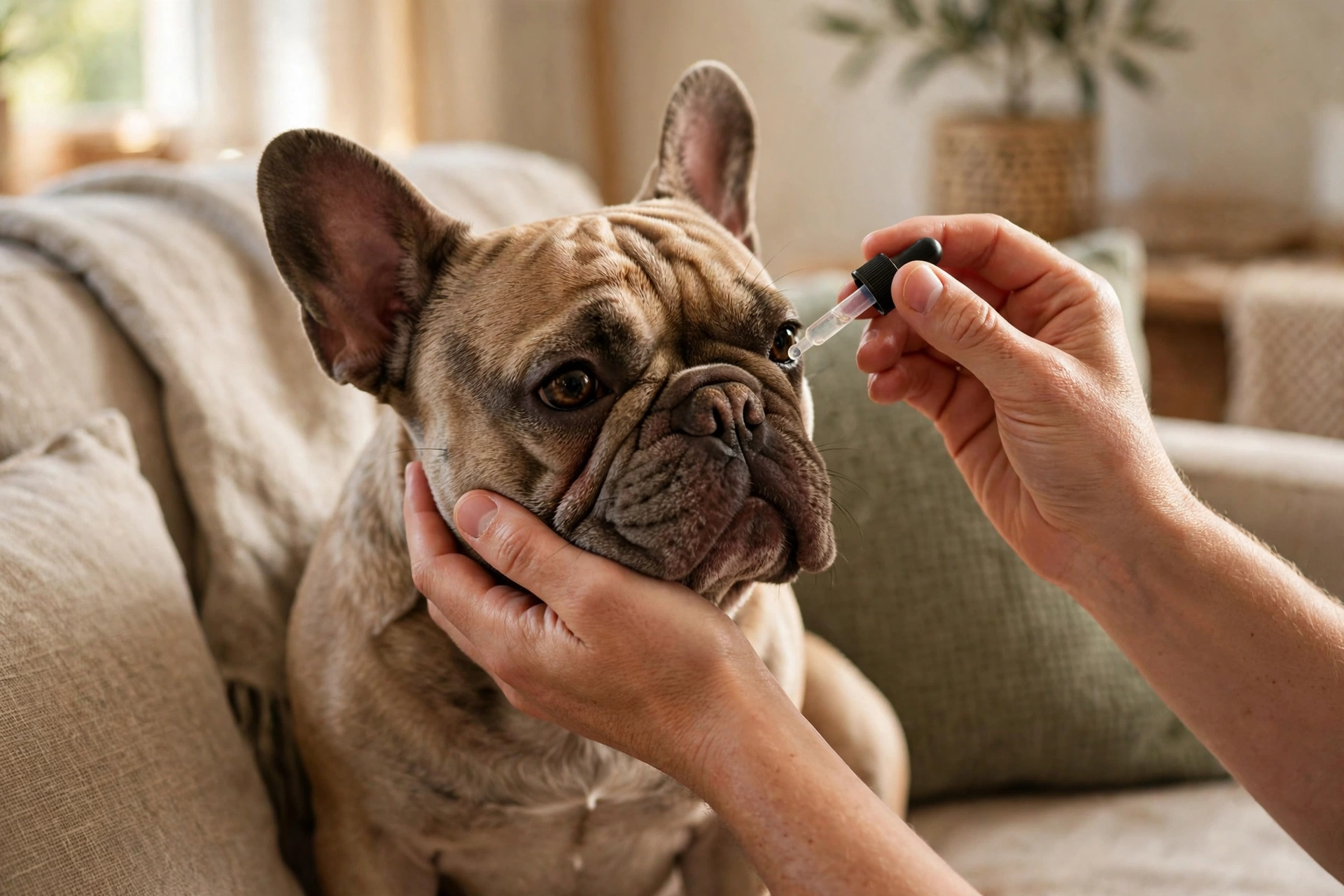
- Do use only what your vet advises for lubrication. Artificial tears ointment can keep the eye moist until surgery. Do not use leftover human eye drops, or anything containing steroids, unless prescribed.
Treatment: Why Surgery Means Tucking, Not Removing
Surgical repositioning of the gland is the gold standard. The American College of Veterinary Ophthalmologists states that to correct prolapse, surgical replacement of the gland is necessary, and that the gland should be repositioned so it can keep functioning normally. VCA’s guidance matches. Treatment involves surgical replacement of the third eyelid gland, and it is essential to treat the condition as soon as possible. The AKC’s veterinary ophthalmologists add the encouraging part. When surgery is performed promptly in a younger dog, it is almost 100 percent effective.
Surgeons use two main techniques. The pocket technique, also called Morgan’s pocket, tucks the gland into a small pocket created in the lining of the third eyelid and stitches it closed. It is the most common approach and the ACVO’s typical procedure of choice. The anchoring or tacking technique stitches the gland to deeper tissue around the eye, sometimes combined with the pocket method in bulldog breeds. An evidence review found no proof that one technique is clearly better, so the surgeon chooses based on the individual dog.
Now the part that matters most. Cutting the gland out was once common, and outdated advice still circulates. Every major authority now advises against removal. About 50 percent of dogs that lose the gland develop dry eye later, compared with about 14 percent after replacement surgery. The ACVO warns that dogs without the gland are predisposed to dry eye later in life, a painful condition that requires the owner to give eye medication multiple times a day for the rest of the dog’s life. Lifelong dry-eye medication runs roughly $30 to $60 a month, on top of ongoing vet monitoring. Removal turns a one-time fix into a lifelong medicated condition. Replacement preserves the gland and succeeds roughly 9 times out of 10.
Checklist: questions for the surgery consult
- Which technique do you recommend for my dog, and why?
- How many cherry eye surgeries do you perform each year?
- What is your re-prolapse rate?
- Will you measure tear production before surgery?
- What does aftercare look like, and exactly what is included in the quote?
Cherry eye is one of several conditions this breed is prone to. Our French Bulldog health problems hub gathers the most common ones in a single place.
Cherry Eye in French Bulldogs: Surgery Costs in 2026
Cost decides timing for most families, so here are the real 2026 numbers. These are ranges from published US cost guides, not a national fee survey. They swing with region, with whether a general practice vet or a specialist operates, and with whether one eye or both need repair.
| Scenario | Typical 2026 US range |
|---|---|
| Per eye, general practice vet | $500 to $1,500 |
| Per eye, board-certified specialist | $1,200 to $2,500 |
| Both eyes repaired in one session | $800 to $2,500 |
| Revision surgery | $400 to $1,000 |
| Pre-op bloodwork, anesthesia, post-op meds | Add $250 to $400 |
The plain-English version. Most US owners pay roughly $500 to $1,500 per eye at a general practice vet in 2026, or $1,200 to $2,500 at a specialist or ophthalmologist. Fixing both eyes in one session typically runs $800 to $2,500 total, often cheaper than two separate operations. Regional gaps are wide. Published examples show West Coast clinics near $1,300 per eye while some East Coast and Southern practices come in around $355.
What pushes the price up: a board-certified ophthalmologist, who typically charges 20 to 30 percent more than a general practice with lower reported recurrence, both eyes in one session, and your zip code. Read the quote carefully. Make sure it includes pre-op bloodwork, anesthesia, and post-op medications, or budget a few hundred more for those.
On insurance: cherry eye is typically treated as pre-existing if it was noted in the records before you enrolled, so early enrollment matters. Revision surgery under an active policy is usually covered.
Quick tip: If the quote stings, ask your vet for a referral estimate from the nearest veterinary teaching hospital. Bundled surgical prices there are often the lowest available, and residents operate under specialist supervision.
One anesthesia note. Cherry eye repair needs general anesthesia, and flat-faced breeds deserve a team that plans for it carefully. Our BOAS guide walks through what the latest anesthesia research found for brachycephalic dogs and which questions to ask any surgical team.
What Happens If You Do Nothing
Waiting is tempting, especially when the dog seems unbothered. The research says waiting is the expensive choice.
An exposed gland dries out and inflames. Tear production falls. About 43 percent of untreated cases develop dry eye. Chronic inflammation takes hold, infections recur, and the cornea can suffer damage. Cornell’s ophthalmology team notes that a chronically prolapsed gland grows very inflamed with persistent discharge, can abrade the surface of the eye, and left untreated can lead to permanent scarring. The ACVO adds the practical warning. The longer the gland stays exposed, the more inflamed it becomes, and a chronic prolapse brings more complications at surgery time. Delay makes the eventual repair harder and less successful.
Quick tip: If money is the reason for waiting, say so at the appointment. Vets navigate this daily and can discuss staged options, teaching-hospital referrals, and payment plans. Silence just reads as not worried.
Recovery: The First Two Weeks and Beyond
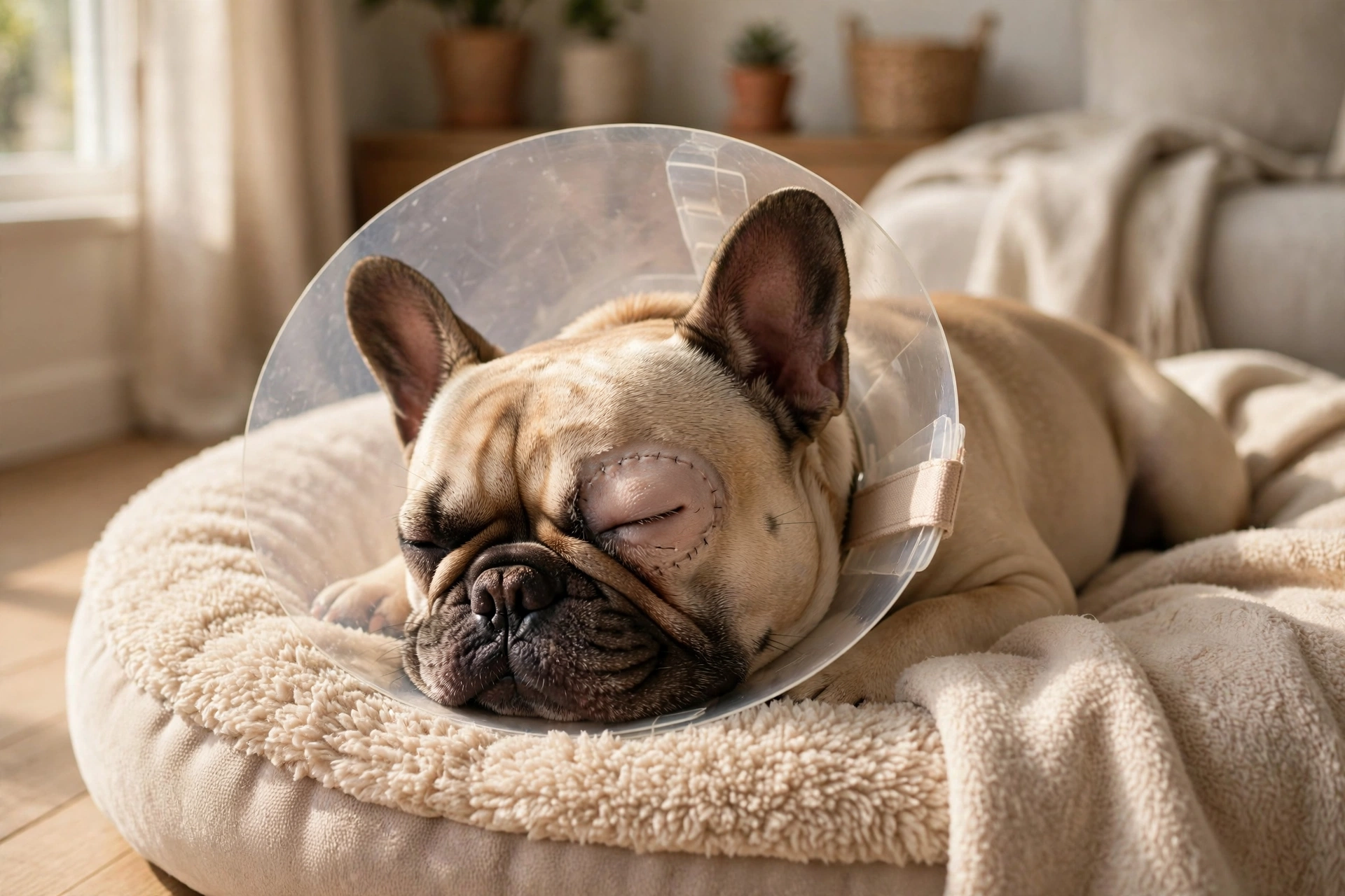
Recovery is straightforward, with one non-negotiable rule. The cone. Your Frenchie wears an e-collar for about two weeks, because the fastest way to undo the surgery is a paw to the eye. Expect eye drops or ointment two to three times a day for two to three weeks, usually alongside oral pain relief and antibiotic eye medication.
The timeline runs like this. The surgical site heals in 10 to 14 days. Your vet will want a recheck at 2 to 4 weeks. Full tissue healing takes 6 to 8 weeks, though most dogs are comfortable within days, and VCA notes the gland typically looks normal again within a few weeks. Across the first months, watch for the gland creeping back out. That is the re-prolapse window.
Keeping a young Frenchie calm for two weeks is the true challenge. Short leash walks replace free play. Puzzle feeders and frozen Kongs replace wrestling. No dog park, no roughhousing, no face-rubbing on the couch cushions.
Recovery kit checklist:
- A properly fitted e-collar. Measure the neck, because a loose cone is a useless cone.
- All prescribed drops and ointments, with a written schedule on the fridge.
- Oral pain medication, given exactly as directed.
- A quiet recovery room away from other pets.
- Your vet’s after-hours number where you can find it at midnight.
Will It Come Back?
The honest answer is that it can. VCA reports that approximately 5 to 20 percent of patients experience a re-prolapse and need another surgery. The AKC’s quoted ophthalmologist puts it at 5 to 10 percent, and notes that a second surgery often uses a different technique. The ACVO cites about 92 percent success, and one study of 420 eyes found 95 percent stayed in place. Bulldog breeds land on the more recurrence-prone end of that range.
When it does return, a revision surgery usually resolves it, typically costing $400 to $1,000. Keep watching the other eye too. VCA notes that many pets with a prolapse in one eye will eventually develop one in the other, with roughly a 50 percent chance tied to the same weak connective tissue on both sides. Some surgeons raise the second eye during the same consult. That call belongs to you and your vet together.
Quick tip: Photograph the eye weekly during recovery, same angle and same light. If the gland starts creeping out again, dated photos show your vet exactly when it began.
When It Is Urgent vs. Routine
Cherry eye on its own is not a midnight emergency. One veterinary site phrases it well. It isn’t an emergency in the middle-of-the-night sense, but it does need a vet. VCA advises bringing any eye mass to your veterinarian’s attention immediately, which means promptly, not eventually.
The lump alone earns a regular appointment soon. Move faster, same day or urgent care, for squinting, a cloudy or bluish eye, thick yellow-green discharge, or bleeding from rubbing. Those signs can mean a corneal ulcer or another problem traveling with the cherry eye, and that is the genuinely urgent part.
Ask for a referral to a veterinary ophthalmologist if the gland has been out for months, if it will not stay down after a vet’s manual attempt, if it re-prolapses after surgery, or if your regular vet seems uncertain. Those are the AKC’s own referral triggers, and requesting one is never an insult.
Do this, not that:
- Do treat the lump as a this-week vet visit. Do not file it under cosmetic and forget it.
- Do mention squinting, cloudiness, or discharge when you call. Do not downplay new symptoms to land a later slot.
- Do ask about a specialist referral when something feels off. Do not assume your vet will take offense. Good vets refer.
Frequently Asked Questions
Does cherry eye hurt my dog? Usually not at first. The lump itself is typically painless, which is why dogs often ignore it until rubbing starts. Squinting or a cloudy eye means a complication has arrived, and that needs prompt attention.
Can both eyes get cherry eye? Yes. It can begin in one eye or both, and roughly half of dogs affected in one eye eventually develop it in the other. Keep watching the unaffected eye during recovery and after.
Is my puppy too young for surgery? No. Cherry eye is a young dog’s disease, with the median first diagnosis under eight months, and vets routinely operate on puppies. Earlier surgery in a young dog carries the best reported outcomes.
Will pet insurance cover cherry eye surgery? Usually yes, provided the condition was not in your dog’s records before enrollment. A lump already documented will likely count as pre-existing. Enroll puppies early, before any eye issue reaches the notes.
Can cherry eye wait a month or two? It is not an emergency, but delay costs. The longer the gland stays exposed, the more inflamed it grows, and chronic cases bring more surgical complications plus higher dry-eye risk. Sooner is genuinely better here.
Why does the lump sometimes vanish and return? Early on, the gland can pop in and out as the loose attachment shifts. That is movement, not healing. It almost always becomes permanently prolapsed without treatment.
Final Word
Cherry eye in French Bulldogs looks alarming but sits firmly in the fixable column. A smooth red lump at the inner corner, a vet visit this week, a tucking surgery that preserves the tear gland, and a couple of cone-wearing weeks at home. The dogs that fare best belong to owners who act early, skip the home massage videos, and refuse gland removal. Photograph the eye today for a baseline, book the appointment, and bring every question from the consult checklist. Your Frenchie will forgive the cone. Save this guide for the recovery weeks, and pass it to the next Frenchie owner who sends you a panicked eye photo at midnight.